Medical history:
Male, 78 y/o pain in right leg. A tibial tumor was diagnosed. Calcemia 13.3; P 2.3; BUN 24; Create 1.6; D vitamin 10.5; PSA 2.5; PTH 1118, compatible with hyperparathyroidism.
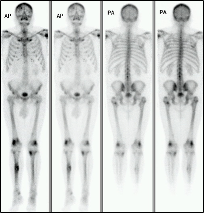
Three-phase bone scan showed intense osteoblastic activity at the middle third of the right tibia and upper third of the contralateral, the lower trochanter and the medial epicondyle of the left femur, lateral epicondyles of both femurs. Also, at the level of kneecaps bilaterally. In addition, a slight and diffuse increase in the uptake at the level of the calvaria, shoulders, sternoclavicular and sacral-iliac joints. There is a small focus of moderate uptake in the lower left maxilla.
Multiple bone lesions were interpreted as metastasis vs hyperparathyroidism with brown tumors.
Echo neck 07/18: Right thyroid cystic solid injury 6.3 x 3.8 x 3.2
FNAP 08/18: Negative cytological for malignant neoplastic cells. Chronic thyroiditis Bethesda group II.
Lab 08/18: TG 18 (N); Ac anti TG <10; TSH 1.75; Free T4 1.15
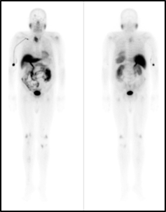
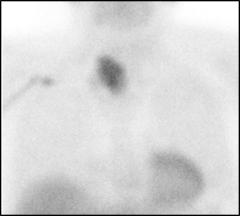
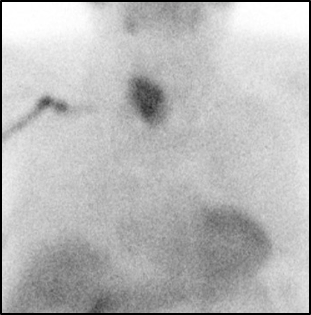
Parathyroid 09/18, showing mass in the right lobe of the thyroid, compatible with hyperfunctioning parathyroid tissue. The bone alterations described can be explained by brown tumors, without being able to rule out secondary lesions. Bone densitometry: osteoporosis of the three locations studied, higher in the right femoral neck.
Surgery was scheduled: parathyroidectomy plus bone biopsy in the right leg. Parathyroid neoplasia of 5x4.5cm with focal capsular involvement. Sample of bone compatible with brown tumors.
Due to clinical and laboratory stability, discharge is decided, with the following diagnoses:
- Primary hyperparathyroidism
- Parathyroid carcinoma
- Brown tumors.
References:
1) Chen Z1 et al. 99mTc-MIBI single photon emission computed tomography/computed tomography for the incidental detection of rare parathyroid carcinoma. Medicine (Baltimore). 2018 Oct;97(40): e12578. doi: 10.1097/MD.0000000000012578.
2) Alabed YZ1 et al. Recurrent parathyroid carcinoma appearing as FDG negative but MIBI positive. Clin Nucl Med. 2014 Jul;39(7): e362-4. doi: 10.1097/RLU.0000000000000357.
3) Désirée Deandreis et al. 18Fluorocholine PET/CT in parathyroid carcinoma: a new tool for disease staging? European Journal of Nuclear Medicine and Molecular ImagingNovember 2015, Volume 42, Issue 12, pp 1941–1942
Home Index Parathyroid Scintigraphy Clinical
Applications |


