Horseshoe kidney
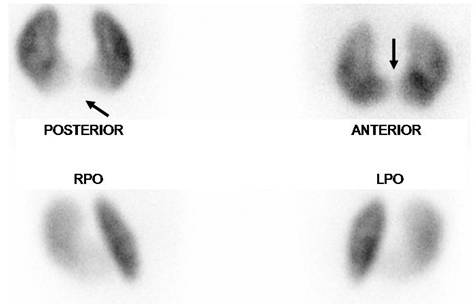
Girl 10 month old. Horseshoe kidney seen on DMSA scan, note homogeneous uptake in both segments, save midline in posterior view due to spine attenuation, corrected in the anterior position (arrows). Relative function is normal, Right segment= 51% and Left segment= 49%. RPO and LPO, right and left posterior obliques.
Horseshoe kidney is easily seen on DMSA scan and it is good for parenchymal assessment.
Reference:
1) Hosokawa S, Kawamura J, Tomoyoshi T, Yoshida O. Congenital renal anomaly: evaluation with 99mTc-dimercaptosuccinic acid renal scintigraphy. Am J Kidney Dis. 1983 May; 2(6): 655-9. 2) Kao PF, Sheih CP, Tsui KH, Tsai MF, Tzen KY. The 99mTc-DMSA renal scan and 99mTc-DTPA diuretic renogram in children and adolescents with incidental diagnosis of horseshoe kidney. Nucl Med Commun. 2003 May; 24(5): 525-30.