Renal Infarct
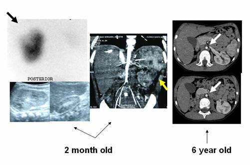
Boy, evaluated at 2 months and at 6 years of age, bearing Takayasu's disease and arterial hypertension. Note diminished aorta lumen (yellow arrow) on angio CT, small right kidney (underwent nephrectomy later on) and enlarged left kidney with a wedge shape defect on ultrasound, better seen in DMSA scan (black arrow). Relative function was 91% in the left side and 9% in the right side. At 6 years of age angio CT (transaxial slices, white arrows) demonstrate small aorta lumen, 2 renal arteries and areas of cortical kidney ischemia.
Renal Scintigraphy is very
sensitive to detect parenchymal defects and infarcts, over 90 %. Although,
it is non specific, the shape with vertex towards the hilum are of help plus the
clinical data. In this case the cause is stenosis of the arteries. Kanat
et al., emphasize the value of DMSA scan for early diagnosis, follow-up of
acute/recurrent renal parenchymal scarring in patients with thrombophilia.
References:
Kanat NB, Aslan M, Bozkurt MF,
Ergün EL. The role of Tc-99m dimercaptosuccinic acid renal cortical scintigraphy
in acute and recurrent episodes of renal infarction in a patient with a tendency
toward thrombosis.Clin Nucl Med. 2009 Oct;34(10):727-30.